


Need the perfect caption for your sunny photos? This guide shares 100+ summer Instagram captions for cute...
A coughing or struggling sheep needs more than a warm corner and crossed fingers. Learn three practical,...
Healthy peach desserts are the sweet spot between fresh summer flavor and everyday balance. This guide shows...
A scissor arm pharmacy wall light blends vintage charm with modern function. This in-depth guide explains what...
A dead battery can turn an ordinary day into a roadside comedy, but hooking up jumper cables...
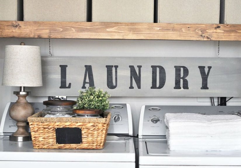
Laundry rooms do not have to be dull, cramped, or purely utilitarian. From small laundry closets and...

A seashell driveway can turn an ordinary entrance into a bright, coastal, well-draining feature that feels both...

Disney’s live-action Hercules remake has no confirmed cast yet, but fans are already building their dream lineup....

Dreaming of a kitchen that feels warm, collected, and effortlessly inviting? These 21 Spanish-style kitchen ideas bring...

What simple thing do you love most? From fresh sheets and morning coffee to kind texts, quiet...