Table of Contents >> Show >> Hide
- What Is a Fluorescein Eye Stain Test?
- Why Doctors Use It: What the Test Can Detect
- How the Test Works (The “Glow” Science, Minus the Boring)
- What Happens During the Fluorescein Eye Stain Test?
- Does It Hurt? Sensations, Side Effects, and Risks
- Understanding Results: What “Positive Staining” Might Mean
- What Happens After the Test?
- When Eye Symptoms Are an Emergency
- FAQ: Quick Answers People Actually Want
- Real-World Experiences (What People Commonly Notice)
- References (Information Synthesized From)
- SEO Tags
If you’ve ever walked into an eye clinic with a scratchy, watery, “something-is-in-there” feeling, there’s a decent chance you met the fluorescein eye stain test.
It’s quick, common, anddepending on the lightingmakes your eyeball look like it belongs in a sci-fi movie (the friendly kind, not the alien abduction kind).
This article breaks down what the fluorescein eye stain test is, why eye-care pros use it, what it can reveal, what the experience feels like,
and what typically happens next. Educational onlynot a substitute for medical care.
What Is a Fluorescein Eye Stain Test?
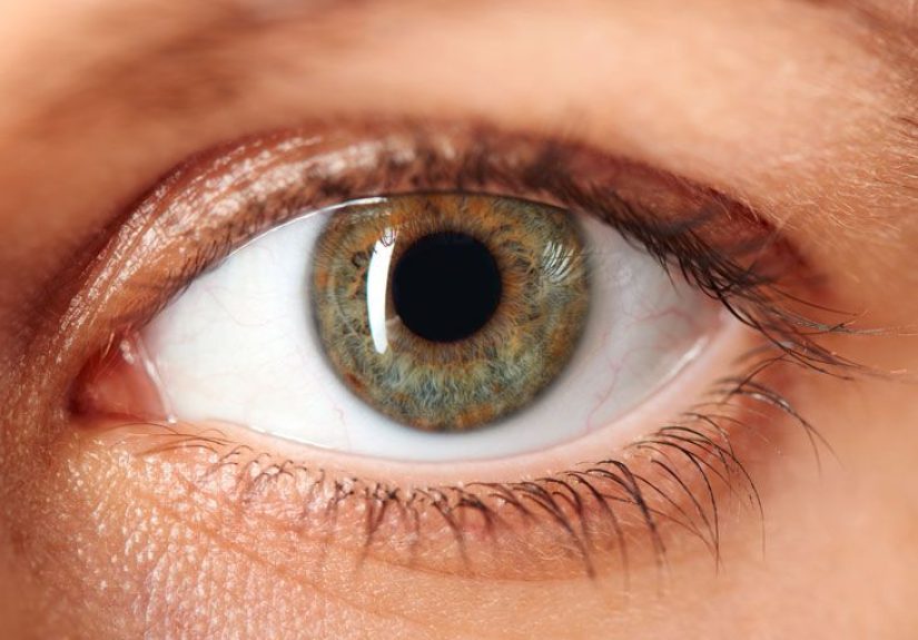
A fluorescein eye stain test (sometimes called a “fluorescent eye test”) is a simple exam that uses a special dyefluoresceinand a blue light
to help a clinician spot problems on the surface of your eye, especially the cornea (the clear “window” at the front of the eye).
Fluorescein is typically a bright yellow-orange dye. Under a cobalt-blue light (often from a slit lamp microscope or a Wood’s lamp),
the dye fluoresces (glows) a vivid green. Areas where the corneal surface is disruptedlike scratchesstand out clearly.
Translation: the test helps your provider “see” tiny surface injuries and patterns that can be hard to spot in normal lighting.
Why Doctors Use It: What the Test Can Detect
The fluorescein eye stain test is used when a provider suspects the front surface of the eye has been irritated, injured, infected, or dried out.
It can help detect (or support the diagnosis of):
- Corneal abrasions (scratches on the cornea), including from fingernails, makeup tools, plant material, sports, or “mysterious pillow-related incidents.”
- Foreign bodies (tiny particles on the eye surface or trapped under the eyelid), like dust, sand, or metal flecks.
- Corneal ulcers and other corneal surface defects that may need urgent evaluation.
- Contact lens–related irritation or surface damage.
- Dry eye–related surface staining (tiny “punctate” staining patterns can show surface stress when tears aren’t doing their job well).
It’s also commonly paired with a close look using a slit lamp microscope and sometimes eyelid eversion (your provider gently flips the eyelid to check underneath).
That sounds dramatic, but it’s usually fast and more “weird” than painful.
How the Test Works (The “Glow” Science, Minus the Boring)
The cornea has layers. When the top surface (epithelium) is intact, fluorescein generally won’t “pool” in a way that screams for attention.
But when there’s a break, the dye collects in those areas, and the blue light makes the defect glow green.
Providers also pay attention to patterns. The shape and distribution of staining can hint at different causes:
a simple abrasion, dryness-related spots, irritation from contact lenses, or more complex corneal problems that need prompt treatment.
What Happens During the Fluorescein Eye Stain Test?
1) Quick questions and a basic eye check
You’ll usually be asked about symptoms (pain, gritty feeling, tearing, light sensitivity), what happened (if anything),
contact lens use, and whether anything got in your eye (especially metal, chemicals, or plant debris).
2) Contacts out (usually)
If you wear contact lenses, your provider typically has you remove them. Fluorescein can temporarily stain soft contacts,
and contacts can complicate both the exam and the interpretation.
3) Dye goes indrop or strip
The dye is applied either as a drop or via a small paper strip moistened with sterile saline. The strip lightly touches the inside of the lower eyelid,
and you blink to spread the dye over the eye surface. It’s quickthink “tap and blink,” not “marinade your eyeball.”
4) Blue light exam (slit lamp or Wood’s lamp)
Your provider uses a cobalt-blue light (often through a slit lamp microscope) to examine the cornea.
Any surface defects can fluoresce bright green. The clinician may also check under the eyelids for trapped debris.
5) Total time: usually minutes
In many clinics or urgent care settings, the entire staining portion takes only a few minuteslong enough for the dye to spread,
the eye to be examined, and the results to be discussed.
Does It Hurt? Sensations, Side Effects, and Risks
Most people tolerate the fluorescein eye stain test well. Common sensations include:
- Mild stinging for a few seconds when drops or dye touch the eye
- Watery eyes (your eyes may tear moreyour body’s “rinse cycle”)
- Temporary yellow-orange discoloration of tears and the skin immediately around the eye if tears spill (hello, dramatic mascara streaks)
Serious reactions from topical fluorescein (the kind used for staining the eye surface) are uncommon. If you’ve had allergies to dyes or eye drops before,
mention it. And if you notice sudden swelling, widespread rash, or breathing trouble after any medication or drop, seek urgent care immediately.
One more important nuance: some people read about dramatic side effects of fluorescein and get worried.
Many of the more systemic reactions (like nausea or more severe allergic responses) are most often discussed in the context of intravenous fluorescein
used for retinal imaging (fluorescein angiography), not the tiny amount placed on the eye surface for a stain test.
Understanding Results: What “Positive Staining” Might Mean
“Positive” in this context generally means the dye highlighted an abnormalityan area of disruption, irritation, or damage on the cornea or conjunctiva.
But the meaning depends on what the staining looks like and the rest of the exam.
Corneal abrasion (scratch)
Abrasions often show up as a bright green area where the cornea’s surface layer is interrupted. The size, shape, and location matter.
For example, linear patterns can suggest something repeatedly rubbing the cornealike a trapped particle under the eyelid.
Foreign body or rust ring concern
If something is embedded or was recently removed, staining can outline the affected area. Metal foreign bodies can leave a rust ring that needs careful evaluation.
This is one reason eye clinicians take “I was grinding metal and now my eye hurts” very seriously.
Dry eye staining patterns
Dry eye can show scattered pinpoint staining (“punctate” staining), often in characteristic regions.
Clinicians may combine staining with other dry eye assessments (tear breakup time, tear production tests, lid margin evaluation)
to decide what type of dry eye is most likely and how to treat it.
Infectious or inflammatory corneal issues
Ulcers, keratitis, or other corneal problems can show more concerning staining patterns and may require prompt treatment and follow-up,
especially for contact lens wearers (who can be at higher risk for serious infections).
Bottom line: the stain test is a powerful “flashlight” for surface problems, but diagnosis is still based on the full clinical picture.
What Happens After the Test?
What comes next depends entirely on what the clinician finds. Common next steps may include:
- Eye irrigation if there’s concern about irritants or small debris
- Foreign body removal (if visible and appropriate to remove in that setting)
- Medication recommendations such as lubricating drops/ointments, and sometimes antibiotic drops/ointment if an abrasion is present
- Contact lens guidance (often: pause contact lens wear until fully healed, depending on diagnosis)
- Referral to ophthalmology for deeper injuries, suspected ulcers, vision changes, severe pain, or high-risk situations
If you’re sent home after the exam, you’ll typically be told what warning signs should prompt urgent reevaluation.
When Eye Symptoms Are an Emergency
Some eye problems can become serious quickly. Seek urgent evaluation (same day, often immediately) if you have:
- Sudden vision loss or significantly blurred vision
- Severe eye pain, especially with light sensitivity
- Chemical exposure (cleaners, solvents, pool chemicals)rinse immediately and get emergency care
- Eye injury from metal, high-speed debris, or a sharp object
- Contact lens wearer + significant pain/redness/discharge
- Obvious swelling around the eye or fever with eye symptoms
The fluorescein stain test is often used in these scenarios because it quickly helps confirm whether the corneal surface is damaged.
But don’t wait for a test if the symptoms are severeget care.
FAQ: Quick Answers People Actually Want
How long does the dye last?
Usually not long. Excess dye rinses out with blinking and tearing. You may notice yellow tears for a short time.
Can I drive afterward?
Many people can, but it depends on whether your provider used other drops (like dilation drops) and how irritated your eye is.
If your vision feels blurry, ask for guidance and consider having someone else drive.
Will it stain my skin or clothes?
The dye can temporarily discolor tears and the skin right around the eye if it runs. It typically washes off skin, but it can stain fabricso maybe don’t wear your favorite white hoodie.
Can I wear contacts right after?
If you came in for pain, redness, or suspected corneal injury, many clinicians recommend pausing contact use until the eye heals.
Follow the specific guidance you’re given.
Is it safe during pregnancy?
Topical fluorescein is widely used, but pregnancy-specific decisions should be individualized.
Tell your provider if you’re pregnant or nursing so they can choose the safest approach for your situation.
Real-World Experiences (What People Commonly Notice)
People often search this test because they’re anxious: eye pain is scary, and it’s hard to “ignore” something that’s literally in the middle of your face.
While everyone’s experience is a little different, there are some recurring themes that show up again and again in patient stories and clinic conversations.
First: the surprise factor. A lot of people expect the test to feel intenselike someone is going to scrub their eyeball with a brillo pad.
In reality, most describe it as more “odd” than painful. The dye strip touch is usually quick, and the stingingif it happensis brief.
Many patients say the hardest part is simply holding still and not blinking on command (because the moment someone says “don’t blink,” your eyelids suddenly become rebellious).
Second: the glow is weirdly reassuring. When the clinician shines the blue light and explains what they’re seeing,
it can feel like the mystery finally has a shape. Patients often report relief when they hear, “Yes, there’s a scratch,” because it turns vague discomfort into something concreteand treatable.
On the flip side, if the provider says, “I don’t see staining,” that can also be reassuring, because it suggests the corneal surface may be intact and the cause could be something less alarming (like dryness or irritation).
Third: watery, neon tears are a whole vibe. People are frequently caught off guard by how much their eye waters afterward.
It’s normal for tears to wash out the dye, and for a short time those tears can look yellowish. Some folks notice a faint yellow tint at the corner of the eye or on the lower lid skin
which is harmless but can look dramatic if you’re wearing makeup. Practical takeaway: tissues are your friend, and rubbing is not.
Fourth: contact lens wearers learn a lesson the hard way. Many contact lens users come in thinking, “It’s just a little irritation.”
When the stain reveals a scratch or a suspicious area, they often hear the same advice: give your eyes a break, treat the surface, and don’t rush back into lenses.
Patients frequently describe frustration herebecause glasses can feel inconvenientbut also admit that a few days of inconvenience beats prolonging healing.
Fifth: the follow-up matters more than the test itself. The stain test is the spotlight; the plan afterward is the real headline.
People who do best tend to follow the clinician’s instructions, return if symptoms worsen, and take warning signs seriously (especially if there’s increasing pain,
worsening redness, sensitivity to light, or any vision change). If you take one “experience-based” truth from this section, make it this:
the test is fast, but your eye’s healing timeline is personallisten to your provider, and don’t try to out-stubborn a cornea.
References (Information Synthesized From)
This article was informed by educational materials and clinical overviews from reputable U.S. health and medical organizations, including:
Healthline; MedlinePlus (U.S. National Library of Medicine); American Academy of Ophthalmology; Cleveland Clinic; American Academy of Family Physicians;
University of Iowa Ophthalmology (EyeRounds/eye atlas resources); Johns Hopkins Medicine; UPMC; WebMD; and NCBI Bookshelf/StatPearls.