Table of Contents >> Show >> Hide
- What Diabetic Neuropathy Is (and Why It’s Hard to Photograph)
- What “Pictures of Diabetic Neuropathy” Usually Show
- A Photo Checklist: What to Look for in Your Own Feet
- Common “Neuropathy Picture” Scenarios (and What Else They Could Be)
- When Pictures Should Trigger a Same-Day Call
- How Clinicians Confirm Diabetic Neuropathy (Beyond Photos)
- What Helps: Prevention and Treatment That Actually Matters
- How to Use Pictures the Right Way (Without Spiraling)
- Conclusion
- Real-World Experiences: What People Commonly Go Through (and What They Wish They Knew Sooner)
If you’ve ever typed “pictures of diabetic neuropathy” into a search bar, you’re not alone. People look for images because symptoms can feel weirdly abstract:
burning, tingling, numbness, “my socks feel bunched up but they’re not.” The catch? Neuropathy is nerve damage, and nerves don’t exactly pose for the camera.
So most “diabetic neuropathy pictures” online aren’t photos of the nerve damage itselfthey’re photos of what can happen because you can’t feel injuries,
or because diabetes affects circulation, skin, joints, and healing.
This guide will help you understand what photos can (and can’t) show, what warning signs tend to appear in real-world images, and how to use pictures responsibly
as a nudge toward care, not a do-it-yourself diagnosis. We’ll keep it clear, practical, and only mildly sarcastic (because your feet deserve respect).
What Diabetic Neuropathy Is (and Why It’s Hard to Photograph)
Diabetic neuropathy is nerve damage linked to diabetes. Over time, high blood sugar can injure nerves and the small blood vessels that supply them,
leading to symptoms that often start in the feet and legs. Neuropathy can be painful, numb, or bothan unfair combo that makes it easy to miss cuts, blisters,
pressure points, and burns.
The main types (because “neuropathy” is a whole family, not one person)
- Peripheral neuropathy: most common; typically affects feet and legs first, sometimes hands.
- Autonomic neuropathy: affects “automatic” body functions (blood pressure changes, digestion, sweating, bladder issues, sexual function).
- Proximal neuropathy: pain and weakness in hips, thighs, or buttocksoften sudden and intense.
- Focal neuropathy: affects a single nerve (for example, a wrist, eye, or part of the face), sometimes abruptly.
Photos can’t capture tingling or burning, and they can’t measure sensation loss. What images can show are the visible footprints neuropathy may leave:
skin breakdown, ulcers, infections, deformities, swelling, redness, and wounds that don’t heal the way they should.
What “Pictures of Diabetic Neuropathy” Usually Show
When people share or search images, they’re usually looking at diabetic foot complicationsoften influenced by neuropathy and sometimes also by poor circulation
(peripheral artery disease). Here are the most common things those photos tend to feature.
1) Dry skin, cracking, and calluses
Many images show heels with deep cracks, thick calluses, or rough, flaky skin. Loss of sweat and oil gland function (which can happen in neuropathy) plus pressure points
can make feet look “overworked,” even if you’ve been sitting all day.
Why it matters: cracks and calluses can become entry points for infection, especially when you can’t feel minor trauma.
2) Blisters and pressure sores you didn’t notice forming
Neuropathy can reduce the ability to feel friction and pressure. Pictures often show blisters on toes or the ball of the footsometimes caused by tight shoes,
seams, or walking “a little extra” on a day that didn’t feel like a big deal.
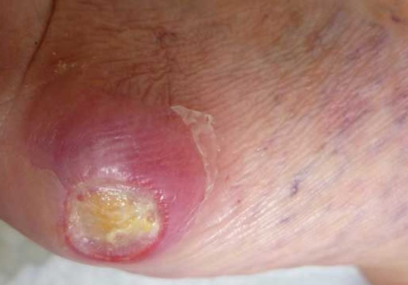
3) Ulcers (open wounds), especially on the sole
A lot of “diabetic neuropathy photos” are actually diabetic foot ulcer images. These can start smalllike a crater under a callusthen deepen.
Some ulcers look like a punched-out hole, others look like a raw red wound, and some are covered by dead tissue.
Why it matters: ulcers can become infected and are a major reason people with diabetes need urgent wound care.
4) Infections (redness, warmth, swelling, drainage)
Photos of infected diabetic wounds often show redness spreading around a sore, swelling, shiny skin, or pus/drainage. Sometimes the skin looks dusky or purple.
Infection can progress quickly, especially when sensation is reduced and treatment is delayed.
5) Charcot foot (swelling + shape changes)
Some of the most dramatic images online are Charcot foot pictures. Charcot changes can begin with redness, warmth, and swellingoften without much pain
and can lead to bone and joint damage and a “rocker-bottom” shape. It can look like a “sudden new foot” showed up overnight (and brought chaos).
6) Toe deformities and muscle wasting
In longer-standing neuropathy, muscle imbalance can contribute to hammertoes or high arches; photos may show toes curling or prominent joints. Some images show thinning
in parts of the foot (atrophy), which can increase pressure pointsbasically turning your foot into its own little stress-test.
A Photo Checklist: What to Look for in Your Own Feet
If you’re using pictures as a self-check tool (smart), aim for consistency: same lighting, same angles, and a routine. A quick daily look is recommended for many
people with diabetesespecially if you’ve had neuropathy symptoms or past foot issues.
Quick daily scan (tops, bottoms, sides, between toes)
- New cuts, blisters, or cracks (even tiny ones)
- Redness, swelling, or warmth compared with the other foot
- Calluses that look thicker, darker, or “bloody” underneath
- Drainage on socks or a spot on bedding (sneaky clue)
- Color changes: pale, blue-ish, purple, or unusually shiny skin
- Toenail changes: thickening, crumbling, or signs of fungal infection
- Any sore that isn’t improving day-to-day
Tip: If bending is tough, use a mirror or your phone camera. You’re not being vainyou’re being preventive.
Common “Neuropathy Picture” Scenarios (and What Else They Could Be)
Photos can help you notice changes, but they can’t tell the full story. Here are examples of what people commonly see in imagesand why you still want clinical guidance.
Scenario A: A dark spot under a callus
Could be: pressure injury, bleeding under the skin, or an ulcer forming under thickened skin.
Also could be: a bruise from minor trauma you didn’t feel, or skin pigmentation changes. If it’s on a pressure point and not improving, treat it as urgent.
Scenario B: Redness around a small cut
Could be: early infection, irritation, or inflammation from rubbing.
Also could be: contact dermatitis or a reaction to a topical product. If redness spreads, warmth increases, or drainage appears, get evaluated quickly.
Scenario C: A swollen, warm foot with little pain
Could be: Charcot changes (especially if swelling is one-sided and persistent).
Also could be: sprain, fracture, gout, or infection. Because neuropathy can blunt pain, “not that painful” doesn’t mean “not serious.”
Scenario D: Numb toes + skin cracks
Could be: peripheral neuropathy plus dry skin issues.
Also could be: vitamin deficiencies, thyroid issues, medication effects, or other causes of neuropathyworth discussing with a clinician.
When Pictures Should Trigger a Same-Day Call
If any of the following show up in a photo (or in real life), don’t “watch and wait” like it’s a questionable TV series. Call your clinician or seek urgent care:
- Spreading redness, significant swelling, warmth, or increasing pain
- Pus/drainage, foul odor, or a wound that looks deeper than it should
- Black or gray tissue (possible dead tissue)
- Fever or feeling unwell along with a foot wound
- Sudden foot shape change or inability to bear weight
- A sore that isn’t clearly improving within 24–48 hours
How Clinicians Confirm Diabetic Neuropathy (Beyond Photos)
Clinicians don’t diagnose neuropathy from pictures alonebecause neuropathy is about nerve function. Common evaluation pieces include:
- Symptom review: burning, tingling, numbness, worse at night, sensitivity to touch.
- Simple sensation tests: a 10-gram monofilament (pressure), vibration testing, temperature or pinprick checks.
- Foot exam: pulses, skin integrity, deformities, calluses, and signs of infection.
- Additional testing when needed: nerve conduction studies or labs to rule out other causes.
A key issue: neuropathy can be asymptomatic in a substantial portion of people with diabetic peripheral neuropathy, which is why routine screening and foot exams matter.
Translation: you can have it even if your feet feel “fine.”
What Helps: Prevention and Treatment That Actually Matters
Managing diabetic neuropathy is usually a two-track plan: (1) slow progression and prevent complications, and (2) treat symptomsespecially painwhen they interfere with life.
Track 1: Protect nerves and prevent foot disasters
- Blood sugar management: steady control helps reduce risk and slow worsening over time.
- Address other risk factors: blood pressure, cholesterol, smoking, and physical activity all matter for circulation and nerve health.
- Daily foot care: wash with warm (not hot) water, dry carefully, moisturize (but not between toes), and check every day.
- Footwear that fits: shoes that don’t rub, socks without harsh seams, and professional help for fitting if you have deformities.
- Regular foot exams: at least yearly for many, more often if you’ve had ulcers, deformities, loss of sensation, or circulation problems.
Track 2: Treat painful neuropathy (without pretending it’s “all in your head”)
Painful diabetic neuropathy can feel like burning, stabbing, electric shocks, or extreme sensitivitysometimes even a bedsheet feels rude. Treatment is individualized, but common
medication categories include certain antidepressants (used for nerve pain), gabapentinoids, and topical options.
In the U.S., there are FDA-approved options specifically for neuropathic pain associated with diabetic peripheral neuropathy, including:
duloxetine, pregabalin, tapentadol extended-release (an opioid option reserved for severe cases when alternatives are inadequate),
and capsaicin 8% patch administered in-office for diabetic nerve pain of the feet.
Non-medication supports can also help: gentle activity, physical therapy for balance and strength, careful sleep strategies, and addressing mood (because chronic pain and depression
love to team up). If pain is severe or function is declining, ask about a referral to a specialist (neurology, endocrinology, podiatry, or pain management depending on the case).
How to Use Pictures the Right Way (Without Spiraling)
Photos are best used for tracking change and catching problems earlynot for diagnosing yourself in a late-night panic scroll.
A simple approach:
- Set a routine: quick daily check; weekly photo set if you’re high-risk or monitoring a specific spot.
- Use good lighting: shadows can turn normal skin into “oh no” skin.
- Compare left vs. right: asymmetry (one foot redder, warmer, more swollen) can be a clue.
- Escalate early: if you’re unsure, send the photo to your clinician’s portal or call. “Small” becomes “big” fast with feet.
Conclusion
“Pictures of diabetic neuropathy” usually means pictures of the visible consequences of nerve damageespecially in the feet: cracks, calluses, blisters, ulcers, infections,
and sometimes Charcot-related shape changes. The most powerful use of photos is not to label what you have, but to notice what’s changing so you can act early.
If you live with diabetes, make foot checks boringly routine. Boring is good. Boring means you keep your toes.
Real-World Experiences: What People Commonly Go Through (and What They Wish They Knew Sooner)
Talk to enough people with diabetes and you’ll hear the same theme: neuropathy rarely announces itself with a marching band. It usually creeps in with “little” sensations that are
easy to dismisstingling at night, a mild burning feeling, toes that feel slightly numb after a long day. Many people describe it as feeling like their feet are wearing invisible socks,
or like they’re standing on pebbles that aren’t actually there. A few shrug it off for months because it’s intermittent, until one day the discomfort becomes loud enough to interrupt
sleep or walking. That’s often when the search for pictures begins: people want something concrete to match what they’re feeling.
One common experience is the “surprise injury.” Someone buys new shoes that feel fine in the store, wears them for a couple hours, and later notices a blisterexcept they didn’t
feel it forming. Or they step on something small (a staple, a splinter, a bit of glass), and only discover it later because they see a tiny blood spot on a sock. Photos become a
record: “This area was normal yesterday; today it’s red.” That timeline can be incredibly useful for clinicians, because diabetic foot problems aren’t just about what the wound
looks likethey’re about how fast it changed.
People also talk about the emotional whiplash of seeing severe images online. It’s easy to stumble into graphic ulcer photos and assume every tingle means catastrophe. The more
grounded reality is that many foot complications are preventable, especially with early detection and consistent care. Folks who do well long-term often adopt simple habits:
a five-minute daily check, moisturizing the right way, trimming nails carefully, breaking in shoes slowly, and getting help for calluses rather than shaving them down at home.
Some say the biggest “unlock” was realizing that foot care isn’t vanityit’s maintenance, like brushing your teeth, except the stakes are higher and your feet can’t text you
when something’s wrong.
Another frequently shared experience: pain that feels out of proportion to what you can see. Someone’s feet may look normal in photos, yet they describe burning that makes
it hard to fall asleep. Others have the oppositeminimal pain but visible swelling or redness that turns out to be serious. This mismatch is exactly why photos are only one tool.
Many people say they wish they’d asked sooner about formal screening tests (like monofilament checks), and they wish they’d known neuropathy can be present even when symptoms
are mild or absent. The “I didn’t know” regret is commonespecially among those who later dealt with ulcers that started as a tiny spot they assumed would heal on its own.
Caregivers often describe a practical challenge: helping someone inspect feet daily without it becoming a battle. The best approach is usually matter-of-fact and routinesame time,
same place, same checklist. Some families take weekly photos to compare changes, especially if there’s a history of ulcers or deformities. The goal isn’t to obsess; it’s to catch
problems early enough that treatment is simpler and outcomes are better. In that sense, pictures can be empowering: not scary trophies from the internet, but a quiet tool for
staying ahead of complications.