Table of Contents >> Show >> Hide
- What Is Superior Vena Cava Syndrome?
- Symptoms of SVCS: What It Can Feel Like (and Look Like)
- What Causes SVCS?
- How SVCS Is Diagnosed
- Treatments for SVCS: What Doctors Actually Do
- Step 1: Supportive care (often started right away)
- Step 2: Endovascular therapy (stents, angioplasty, and clot-directed treatment)
- Step 3: Cancer-directed therapy (chemotherapy and/or radiation)
- Step 4: Anticoagulation (when clots or devices are involved)
- Step 5: Surgery (less common, but still a tool)
- SVCS in Real Life: Two Quick Examples
- Possible Complications (and Why SVCS Gets Attention Fast)
- Living With (or After) SVCS: Practical Takeaways
- Experiences With SVCS: What People Commonly Notice (A 500-Word Add-On)
Your body has a lot of “traffic lanes” for blood. The superior vena cava (SVC) is one of the biggestand it’s basically the expressway
that returns blood from your head, neck, arms, and upper chest back to your heart. When that expressway gets squeezed or blocked,
blood backs up like rush-hour traffic with no alternate route… until your body tries to build detours (collateral veins). That backup creates a group of
signs and symptoms called Superior Vena Cava Syndrome (SVCS).
SVCS can creep in slowly over days or weeks, or it can show up fast and demand urgent careespecially if breathing is affected. The good news: modern
imaging and treatments (including minimally invasive procedures) can often relieve symptoms quickly while doctors treat the underlying cause.
The goal of this guide is to explain SVCS in plain, standard American Englishwith enough depth to be useful, without turning it into a medical textbook
you didn’t ask for.
What Is Superior Vena Cava Syndrome?
Superior vena cava syndrome is not one single disease. It’s a syndromea recognizable patterncaused by
partial or complete obstruction of blood flow through the SVC. The blockage can happen because something outside the vein is pressing
on it (like a tumor or enlarged lymph nodes), or because something inside the vein is narrowing or clogging it (like a blood clot around a catheter or
pacemaker lead).
Think of the SVC as a wide, low-pressure tube. Low pressure is great for efficient blood returnuntil it’s not. When the SVC is compressed or blocked,
pressure rises upstream, and fluid can leak into tissues. That’s why swelling is such a common theme in SVCS.
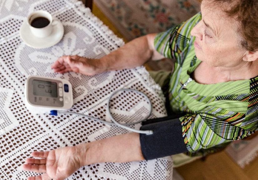
Symptoms of SVCS: What It Can Feel Like (and Look Like)
SVCS symptoms usually show up in the areas that drain into the SVC: the face, neck, arms, and upper chest. Some symptoms are mild and
annoying. Others can be seriousespecially when swelling affects the airway or causes brain-related symptoms due to
impaired venous drainage.
Common symptoms
- Swelling of the face (often worse in the morning or when bending over)
- Neck swelling and a “tight collar” feeling
- Swelling of the arms or hands
- Shortness of breath, especially when lying flat (orthopnea)
- Cough or a sense of chest pressure
- Visible, bulging veins in the neck or on the upper chest
- Hoarseness or trouble swallowing (sometimes)
- Head fullness or facial redness/bluish discoloration
More urgent “red flag” symptoms
SVCS can be considered a medical emergency when symptoms suggest airway compromise, reduced blood return affecting the heart,
or increased pressure affecting the brain. Symptoms that should be taken seriously include:
- Severe trouble breathing or noisy breathing
- Confusion, fainting, or significant dizziness
- Severe headache with worsening facial/neck swelling
- Swelling that progresses quickly over hours
Important note: SVCS symptoms can overlap with other conditions (allergies, infections, heart failure, even “just” a stubborn cough). The difference is
the patternupper-body swelling plus visible veins plus breathing issues often points clinicians toward SVCS faster than a detective in a crime show who
found the murder weapon labeled “MURDER WEAPON.”
What Causes SVCS?
Causes are often grouped into two buckets: malignant (cancer-related) and benign (non-cancer). Historically, cancer
caused most cases. Today, benign cases are increasingly recognized due to the widespread use of central venous catheters,
pacemakers, and other intravascular devices.
1) Cancer-related causes (malignant SVCS)
Malignancy can compress or invade the SVC directly or enlarge nearby lymph nodes that press on it. Common cancer-related causes include:
- Lung cancer (a frequent cause because the SVC sits in the chest near lung structures)
- Non-Hodgkin lymphoma or other mediastinal lymphomas
- Metastatic cancers that involve the chest lymph nodes
2) Non-cancer causes (benign SVCS)
Benign SVCS is often related to a blood clot (thrombosis) or scarring/narrowing of the vein. Causes can include:
- Central venous catheters (including long-term IV lines)
- Pacemaker/ICD leads and other implanted devices
- Blood clots due to clotting disorders (less common)
- Fibrosing mediastinitis (rare scarring condition in the mediastinum)
- Dialysis access-related venous narrowing/thrombosis (in certain situations)
Why the cause matters so much
The cause determines the best next step. If SVCS is from a tumor, treatment usually targets the cancer (chemotherapy, radiation therapy, or both), and
symptom relief may come from an endovascular stent. If SVCS is from a clot around a catheter, doctors may focus on
anticoagulation, catheter management, and sometimes thrombolysis or stenting depending on severity.
How SVCS Is Diagnosed
Diagnosis starts with a careful history and physical exam. Visible chest wall veins, neck vein distention, facial/arm swelling, and breathing symptoms
can strongly suggest SVCS. From there, imaging confirms the diagnosis, identifies the location of obstruction, and helps determine the cause.
Common tests and imaging
-
Contrast-enhanced CT (CT chest with IV contrast): Often the workhorse test. It can show a tumor, enlarged lymph nodes, clot, and
collateral veins. - CT venography or specialized venous-phase imaging: Helps map venous flow patterns and blockages more precisely.
- Catheter venography: Sometimes used when an intervention is planned (it can diagnose and treat in the same session).
- Ultrasound: Can evaluate arm veins for clot, but the SVC itself is deep in the chest and less accessible by ultrasound.
- Biopsy (when cancer is suspected): Identifying the cancer type can guide the fastest, most effective therapy.
One practical point clinicians often consider: if the patient is stable, they may try to establish a diagnosis of the underlying cancer (like lymphoma vs.
lung cancer) before starting treatments that could blur the tissue diagnosis. But if symptoms are severe, the priority shifts to stabilizing and relieving
obstruction quickly.
Treatments for SVCS: What Doctors Actually Do
SVCS treatment has two goals:
(1) relieve the obstruction and symptoms and (2) treat the underlying cause. The exact plan depends on how severe the
symptoms are and whether the cause appears malignant or benign.
Step 1: Supportive care (often started right away)
Supportive measures don’t “cure” SVCS, but they can reduce discomfort and buy time while definitive treatment is organized. In clinical settings, this may include:
- Keeping the head elevated (to reduce venous pressure in the upper body)
- Oxygen if breathing is strained
- Diuretics in selected cases to help reduce fluid overload
- Corticosteroids in specific scenarios (for example, when steroid-responsive tumors are suspected or swelling is significant)
Not every supportive tool fits every patient. The key point is that SVCS is usually managed by a teamoften emergency clinicians, oncologists, pulmonologists,
interventional radiologists, cardiologists, and sometimes surgeonsbecause the “right” move depends on the “why.”
Step 2: Endovascular therapy (stents, angioplasty, and clot-directed treatment)
Over the past couple of decades, endovascular stenting has become a leading option for rapid symptom reliefespecially when SVCS is severe
or when quick palliation is needed. The procedure is typically performed by an interventional radiologist or similar specialist and may involve:
- Balloon angioplasty (temporarily widening the narrowed segment)
- Stent placement (a mesh tube to keep the vein open)
- Thrombolysis (clot-dissolving therapy) or thrombectomy (clot removal) in select clot-driven cases
When it worksand it often doespatients can notice improvement in swelling and breathing relatively quickly. But endovascular therapy is not a magic wand.
Doctors still need to address the root cause, and patients may require medications afterward such as anticoagulants depending on why the
obstruction happened and what devices are involved.
Step 3: Cancer-directed therapy (chemotherapy and/or radiation)
When SVCS is caused by cancer, the long-term solution is treating the cancer. Options may include:
- Chemotherapy: Often used for chemo-sensitive cancers like some lymphomas and small cell lung cancer.
- Radiation therapy: May shrink tumors compressing the SVC and reduce symptoms, sometimes within days to a couple of weeks.
- Immunotherapy/targeted therapy: Depending on tumor type and biomarkers, these may be part of the treatment plan.
In real-world practice, teams may combine approachesfor example, a stent for fast symptom relief plus chemo or radiation to control tumor growth.
The sequence is individualized. The “best” plan is the one that balances speed, safety, and effectiveness for that patient’s diagnosis.
Step 4: Anticoagulation (when clots or devices are involved)
If SVCS is related to thrombosis (a blood clot), clinicians often consider anticoagulationespecially when a catheter,
pacemaker lead, or underlying clotting risk is part of the story. Sometimes the device may be repositioned or removed; sometimes it’s essential and must
stay, and the plan focuses on preventing clot progression and maintaining venous flow.
The details (which anticoagulant, how long, whether thrombolysis is needed) depend on severity, bleeding risk, and the exact anatomy of the clotso this
is a classic “do not try to freestyle it at home” situation.
Step 5: Surgery (less common, but still a tool)
Open surgery is less common today because endovascular therapy is often effective and less invasive. But surgery may be considered for selected patients,
especially when anatomy is complex, when prior treatments failed, or when reconstruction/bypass is needed.
SVCS in Real Life: Two Quick Examples
Example A: Gradual onset with cancer
A 62-year-old develops weeks of worsening facial puffiness, shortness of breath when lying flat, and prominent chest veins. CT imaging shows a chest mass
and enlarged lymph nodes compressing the SVC. The team confirms the cancer type and starts cancer-directed therapy. If symptoms are severe, a venous stent
may be placed to relieve obstruction quickly while treatment begins.
Example B: Device-related clot
A 48-year-old with an implanted device notices arm swelling and neck vein fullness. Imaging shows a clot and narrowing near the SVC associated with leads.
Management may include anticoagulation and, if symptoms are significant or flow is severely limited, endovascular intervention to restore patency.
Same syndrome, different cause, different strategythis is why SVCS is treated like a “find the culprit first” mystery.
Possible Complications (and Why SVCS Gets Attention Fast)
Many people with SVCS mainly experience uncomfortable swelling and shortness of breath. But the condition can become dangerous when it affects:
- The airway (swelling can worsen breathing)
- The brain (increased venous pressure may contribute to neurologic symptoms)
- Circulation (reduced venous return can contribute to hemodynamic stress in severe cases)
Clinicians also watch for complications tied to the underlying causesuch as cancer progression or recurrent thrombosisbecause SVCS is often the “signal”
of a bigger issue that needs treatment.
Living With (or After) SVCS: Practical Takeaways
- SVCS is treatable, and relief can sometimes be rapid with modern interventions.
- Diagnosis matters: cancer-related SVCS and clot/device-related SVCS are managed differently.
- Imaging is key, often with contrast CT to map the obstruction and identify the cause.
- Follow-up is important because the underlying condition (cancer or thrombosis risk) determines long-term outcomes.
If you’re reading this because you or someone you care about might have SVCS: it’s okay to feel rattled. “A blocked vein near the heart” sounds like the
kind of plot twist nobody requested. But SVCS is a well-known clinical problem with established diagnostic pathways and multiple treatment options.
The fastest path to feeling better is usually getting evaluated promptly so the cause can be identified and treated correctly.
Experiences With SVCS: What People Commonly Notice (A 500-Word Add-On)
SVCS has a weird social feature: it’s one of the few medical problems where friends might say, “You look different,” before you even feel seriously ill.
Many people describe a subtle beginningwaking up with a puffy face that looks like a bad allergy day, or noticing rings feel tighter, or a hoodie neckline
suddenly feels like it shrank in the wash. But the hoodie didn’t betray you. Your venous return did.
A common story is the “morning face” pattern. People may notice swelling is worse after lying down and improves somewhat during the day. That can create
a cycle of second-guessing: “Maybe it’s salt. Maybe it’s sleep. Maybe it’s stress.” Then the veins in the neck or upper chest become more visible, and
breathing starts to feel slightly harder when bending over to tie shoes. That combination is often what finally pushes someone to seek care.
For people whose SVCS is related to cancer, the emotional experience can be a double hit: first the fear from symptoms that feel dramatic (swelling, breath
changes), and then the shock of discovering the cause. Many patients say the early days feel like an avalanche of new vocabularyCT, biopsy, staging,
radiation oncology, interventional radiologyplus a parade of specialists. One practical coping strategy people often find helpful is bringing a notebook
(or using a notes app) and writing down three things at each visit:
What we know, what we’re doing next, and what symptoms should trigger urgent help.
For device- or clot-related SVCS, experiences can be different. Some people feel frustrated because they “did everything right”they got the pacemaker or
central line to improve health, and now there’s a complication. In these cases, patients often describe feeling better once there’s a clear plan:
anticoagulation, device assessment, and a timeline for follow-up imaging. The uncertainty tends to be the hardest partso clear explanations and a written
plan can be as calming as the medication itself.
People who undergo stenting often describe the relief as surprisingly fastlike someone “turned the pressure down.” Swelling doesn’t always vanish
instantly (bodies love gradual transitions), but breathing and head fullness may improve sooner than expected. Afterward, many patients say the most useful
guidance is practical: what activity is okay, which symptoms are expected during recovery, how to manage follow-up appointments, and what signs could mean
the vein is narrowing again.
Finally, caregivers often share their own experience: SVCS looks scary. Watching a loved one struggle to breathe or swell rapidly can be alarming. Many
caregivers feel better when clinicians explain that SVCS is a known pattern with a defined workupand that the team’s first priority is stabilizing
breathing and circulation while identifying the cause. If there’s one “experience-based” takeaway, it’s this:
SVCS is serious, but it’s also something healthcare teams are trained to recognize and treat. Getting evaluated quickly is not overreacting.
It’s being smart.