Table of Contents >> Show >> Hide
- What “Stage 1” Really Means (and Why the Details Matter)
- How Mammograms Detect Stage 1 Breast Cancer
- From Screening to Diagnosis: The “Callback” Path (No, It’s Not a Personal Failing)
- Common Mammogram Findings That Lead to Stage 1 Detection (With Real-World Examples)
- After Detection: A Stage 1 Treatment Snapshot (What Often Happens Next)
- Prognosis and Follow-Up Mammograms
- Screening Mammogram Timing: Why the Guidelines Can Sound Confusing
- Smart Questions to Ask After a Mammogram (or a Callback)
- Experiences Related to Stage 1 Breast Cancer and Mammogram Detection
- Conclusion
If breast cancer had a personality, Stage 1 would be the version that tries very hard not to make a scene.
It’s early, often small, and frequently discovered before you’d ever feel a lumpthanks to one of modern medicine’s
least glamorous heroes: the mammogram.
This guide breaks down what Stage 1 breast cancer actually means, how mammograms find it, why you might get a dreaded
“callback,” and what the typical next steps look like. We’ll keep it factual, practical, and just funny enough to
make the waiting room magazines jealous.
What “Stage 1” Really Means (and Why the Details Matter)
“Stage 1 breast cancer” is an early-stage diagnosis. In plain English, that usually means:
the cancer is small and either hasn’t spread to nearby lymph nodes or has only tiny amounts of cancer cells in a nearby node.
Stage 1 is not “nothing,” but it is often very treatable.
Stage 1A vs. Stage 1B: Same neighborhood, different house numbers
Doctors stage breast cancer using the TNM system:
T for tumor size, N for lymph node involvement, and M for distant spread.
Stage 1 typically sits in the “small tumor, limited spread” range.
- Stage 1A: The tumor is usually 2 cm (about 0.8 inches) or smaller and there’s no sign it has spread to lymph nodes.
-
Stage 1B: The tumor may be small (or sometimes not clearly seen as a mass), but there are
tiny clusters of cancer cells in nearby lymph nodes (often called micrometastases).
Why does this matter? Because staging helps predict prognosis and guides treatment choiceslike whether you’ll likely
need radiation, hormone therapy, targeted therapy, chemotherapy, or some combination. Stage 1 isn’t one-size-fits-all;
it’s more like a “choose your own adventure,” except nobody asked for this book.
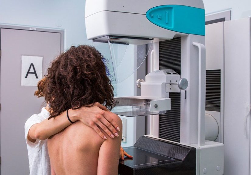
How Mammograms Detect Stage 1 Breast Cancer
A mammogram is a specialized X-ray of the breast designed to spot changes that can be too small to feel.
The big win with mammograms is timing: finding a cancer when it’s small and localized can make treatment simpler and outcomes better.
What a mammogram is actually looking for
Mammograms don’t “see cancer” like a movie villain scanner. They detect patterns and abnormalities that might represent cancer.
Common red flags include:
- Masses (a lump-like area that looks different from surrounding tissue)
- Calcifications (tiny calcium deposits; certain patterns can raise concern)
- Asymmetry (one area doesn’t match the same region in the other breast)
- Architectural distortion (tissue looks “pulled” or warped, even without a clear mass)
Stage 1 cancers are often found because they’re small enough to hide from your fingers but not from a well-read image.
That’s one reason screening matters: it can catch changes before symptoms show up.
2D mammograms vs. 3D mammograms (tomosynthesis)
Many imaging centers now use 3D mammography (digital breast tomosynthesis), which takes multiple images from different angles.
Think of it as flipping through pages instead of staring at the coverhelpful when breast tissue overlaps on a standard 2D view.
In practice, 3D imaging can improve detection of some cancers and may reduce unnecessary callbacks for some people.
The big plot twist: breast density
Breast density refers to the mix of fatty tissue and fibrous/glandular tissue in the breast.
Dense tissue can make it harder to spot cancer on a mammogram because both dense tissue and many tumors appear lighter on X-rays.
Dense breasts are also associated with a higher risk of developing breast cancer.
Since late 2024, mammogram reports in the U.S. generally include a standardized breast density notification (“dense” or “not dense”),
which is meant to help patients have more informed conversations with their clinicians about screening options.
From Screening to Diagnosis: The “Callback” Path (No, It’s Not a Personal Failing)
Let’s talk about the word that makes people’s stomachs drop: callback.
After a screening mammogram, some people are asked to return for additional imaging.
This is common and usually means the radiologist needs a closer looknot that you definitely have cancer.
How common are callbacks?
In the U.S., about 1 in 10 screening mammograms leads to a callback for more testing.
And among people who are called back, most do not end up with a cancer diagnosis.
Callbacks happen because breasts are complicated, shadows happen, and radiologists prefer certainty over guesswork.
What typically happens after a callback
-
Diagnostic mammogram:
More targeted images (extra angles, magnification views) focusing on the area that looked different. -
Breast ultrasound:
Often used to evaluate a mass or clarify whether something is a fluid-filled cyst vs. a solid area. -
Breast MRI (in selected cases):
More common for higher-risk screening, assessing extent of disease, or clarifying complex findings. -
Biopsy:
If imaging remains suspicious, a biopsy removes a small sample for lab testing. This is what confirms diagnosis.
Understanding BI-RADS (the radiology scorecard)
Imaging results are commonly categorized using BI-RADS, a standardized system radiologists use to communicate risk and next steps.
You might see categories like:
- BI-RADS 0: Incompletemore imaging needed (a common “callback” category)
- BI-RADS 1–2: Negative or benign findings
- BI-RADS 3: Probably benignshort-interval follow-up imaging is recommended
- BI-RADS 4–5: Suspicious to highly suspiciousbiopsy is usually recommended
- BI-RADS 6: Known cancer (used after biopsy confirms diagnosis)
Important: BI-RADS is a communication tool, not a final diagnosis. If you see a higher number, it means “let’s verify,” not “we already know.”
Common Mammogram Findings That Lead to Stage 1 Detection (With Real-World Examples)
Stage 1 breast cancer can show up in different ways. Here are a few scenarios that are common in clinical practice:
Example 1: Suspicious calcifications
A routine screening mammogram shows a cluster of tiny calcifications with a pattern that’s not clearly benign.
The callback includes magnification views. If the pattern remains concerning, a stereotactic biopsy
may be recommended. Sometimes this finds very early invasive cancer or a pre-invasive condition (like DCIS) that needs treatment.
Example 2: A small spiculated mass
A screening image reveals a small mass with irregular edges. Ultrasound confirms a solid lesion.
A core needle biopsy is performed, and pathology identifies an invasive cancer. If it’s under 2 cm and nodes are negative,
this can be staged as Stage 1A.
Example 3: Architectural distortion with no obvious lump
Sometimes the tissue looks “tugged” or distorted, even when there’s no clear ball-shaped mass.
3D mammography can be especially helpful here. If follow-up imaging stays suspicious, biopsy may reveal a small cancer that would have been easy to miss otherwise.
After Detection: A Stage 1 Treatment Snapshot (What Often Happens Next)
Treatment plans are personalized based on the tumor’s size, grade, lymph node status, and biomarkers such as hormone receptor status (ER/PR)
and HER2 status. But many Stage 1 treatment paths share a familiar skeleton:
Surgery: Lumpectomy vs. mastectomy
Many Stage 1 cancers are treated with either:
-
Lumpectomy (breast-conserving surgery): removes the tumor with a rim of normal tissue.
It’s often followed by radiation therapy to reduce recurrence risk. -
Mastectomy: removes more breast tissue (sometimes all). This may be recommended based on tumor factors,
multiple areas of disease, genetic risk, prior radiation, or patient preference.
A sentinel lymph node biopsy is commonly performed to check whether cancer cells have reached the first draining lymph nodes.
Stage 1 usually means nodes are negative or only have very tiny deposits.
Radiation therapy
Radiation is commonly recommended after lumpectomy. For early-stage cancers, many people receive whole-breast radiation,
and some may qualify for partial-breast approaches depending on individual factors. The goal is to reduce the chance of cancer returning in the breast.
Systemic therapy: the “insurance policy” treatments
Even when a tumor is small, your care team may recommend treatments that reduce recurrence risk throughout the body.
These decisions often depend on tumor biology:
-
Hormone (endocrine) therapy for hormone receptor-positive cancers (often taken for several years).
This helps lower the risk of recurrence. - HER2-targeted therapy for HER2-positive cancers (sometimes paired with chemotherapy), because HER2-positive tumors can behave more aggressively.
- Chemotherapy for higher-risk situations (for example, certain tumor grades, biomarker patterns, or genomic test results).
Translation: Stage 1 is early, but the treatment plan still aims for “finish the job, lock the doors, and set the alarm.”
Prognosis and Follow-Up Mammograms
Outcomes for early-stage breast cancer are often very good. National registry-based statistics show that
breast cancers found before spreading beyond the breast (localized disease) have a very high 5-year relative survival rate.
That’s one reason detection at Stage 1 matters.
What follow-up imaging can look like
Follow-up varies by treatment and risk, but common patterns include:
- Regular clinical visits with your oncology or surgical team, especially in the first few years
- Annual mammograms of the remaining breast tissue after lumpectomy (and of the other breast)
- Additional imaging (like MRI) for certain higher-risk people, depending on genetics, density, and personal history
A good survivorship plan also covers symptom monitoring, medication side effects (if you’re on endocrine therapy), and lifestyle support.
The point isn’t to live in fearit’s to stay a step ahead with smart, scheduled check-ins.
Screening Mammogram Timing: Why the Guidelines Can Sound Confusing
If you’ve ever Googled “when should I get a mammogram?” and ended up with 14 tabs and a headache, you’re not alone.
Different medical organizations weigh benefits and trade-offs differently (like reducing cancer deaths vs. increasing false positives).
A widely cited baseline for average-risk adults in the U.S. is:
screening mammography every 2 years from ages 40 to 74.
Other professional groups recommend annual screening starting at 40 and emphasize earlier risk assessment
to identify people who may need earlier or supplemental screening.
When your “average risk” isn’t actually average
You may need earlier or more intensive screening if you have factors like:
- A strong family history of breast or ovarian cancer
- A known genetic mutation associated with higher breast cancer risk
- A history of chest radiation at a young age
- Very dense breasts plus additional risk factors
The most useful move is a risk conversation with a clinician who can map your personal factors to a screening plan.
The best guideline is the one that fits you.
Smart Questions to Ask After a Mammogram (or a Callback)
You don’t need a medical degree to advocate for yourself. A few questions can clarify the situation fast:
- What BI-RADS category was assigned, and what does it mean for next steps?
- Is the finding new compared to my prior mammograms?
- Do I have dense breasts, and does that change my screening strategy?
- What type of follow-up imaging is recommended (diagnostic mammogram, ultrasound, MRI), and why?
- If a biopsy is recommended, what kind and what should I expect for timing and results?
Pro tip: if you’ve had prior mammograms at another facility, ask about transferring those images. Comparisons can reduce callbacks and improve interpretation.
Experiences Related to Stage 1 Breast Cancer and Mammogram Detection
Facts and guidelines are helpfulbut people don’t live their lives in bullet points. The mammogram-to-diagnosis path
has a very human soundtrack: waiting, Googling, overthinking, and then trying to remember what day it is.
Below are common experiences people report (shared here in a general, educational wayyour situation can be different).
The mammogram appointment: “That was it?” (and also: “That was it.”)
Many describe the screening mammogram as quicksometimes just minutes once you’re positionedfollowed by an immediate urge
to reward themselves with coffee like they just ran a marathon. The compression can feel uncomfortable, but it’s brief.
What surprises a lot of people isn’t the imagingit’s the emotional weight they didn’t expect to carry in the car ride home.
The callback phone call: a masterclass in sudden anxiety
People often say the hardest part is hearing, “We need additional images.” Even when they’re told it’s common, the brain
tends to respond with: “COMMON FOR WHO, EXACTLY?” It can help to remember that callbacks are frequently about clarity:
a shadow, an overlap of tissue, or a small spot the radiologist wants to magnify. Many callbacks end with reassurance.
Waiting for results: time becomes soup
Waiting is where imaginations go to do cardio. People describe refreshing patient portals, rereading the same sentence 12 times,
and developing a deep personal relationship with their phone’s notification sound. A practical coping trick some share:
schedule something normal afterward (a walk, a movie, dinner with a friend) so your whole day isn’t “The Day of Waiting.”
When the news is Stage 1: relief + fear, at the same time
“Early” can bring a wave of relieffollowed immediately by the realization that it’s still cancer, and you still have decisions to make.
People often say the language matters: they want clinicians who celebrate early detection while also taking their fear seriously.
Many find it grounding to ask for a written plan: what tests are next, what the likely timeline is, and who their main point of contact will be.
Common “I wish I’d known” notes from the early-stage journey
- Bring a notebook (or a friend who takes notes). Appointments can be information-heavy, and it’s easy to forget details under stress.
- Ask for your pathology basics in plain language. Tumor size, grade, hormone receptors, and HER2 status influence treatment decisions.
- Second opinions can be calming, not confrontational. Many people say a second opinion made them feel more confidentregardless of whether the plan changed.
- It’s okay to feel “not sick” and still overwhelmed. Stage 1 can come with few or no symptoms, which can make the diagnosis feel surreal.
- Support can be practical, not just emotional. People often value help with rides, meals, childcare, or just someone to sit quietly with them.
If there’s one recurring theme, it’s this: mammogram detection often finds Stage 1 breast cancer before it “announces itself.”
That early timing can open doors to more options and strong outcomes. But it’s still a lotso treating your emotional experience
as real and valid is not drama. It’s being human.
Conclusion
Stage 1 breast cancer is often discovered through mammogram detectionsometimes before any symptoms show up.
Understanding what Stage 1 means (and how staging works), what mammograms can and can’t see (hello, breast density),
and why callbacks happen can take some of the mystery out of an already stressful process.
If you take away only one thing: a callback is a request for clarity, not a verdictand early detection gives you a powerful head start.
For personalized decisions about screening and treatment, the best next step is a conversation with your healthcare team based on your individual risk and results.