Table of Contents >> Show >> Hide
- What Is an Electron Microscope Calibration Target?
- Why Build a DIY Calibration Target?
- The Best DIY Strategy: Assemble, Don’t Fabricate
- Materials You Can Use for a DIY SEM Calibration Target
- How to Build a Practical DIY Calibration Target
- How to Use Your DIY Calibration Target
- Simple Calibration Math
- Documentation: The Secret Ingredient Nobody Brags About
- What a DIY Calibration Target Cannot Do
- Common Mistakes to Avoid
- DIY Calibration Target Ideas by Budget
- Best Practices for Long-Term Use
- DIY Calibration Target for TEM: A Quick Note
- Real-World Experience: What You Learn After Building One
- Conclusion
A DIY calibration target for electron microscopes sounds like the sort of weekend project that starts with confidence, a cup of coffee, and the dangerous phrase, “How hard could it be?” The honest answer: not impossible, but not casual either. Electron microscopes do not forgive sloppy rulers. At SEM and TEM magnifications, a tiny error in scale can turn a 500-nanometer particle into a 620-nanometer particle, and suddenly your beautiful measurement is wearing clown shoes.
The good news is that a practical DIY calibration target does not require you to fabricate nanoscale lines from scratch in a garage laboratory. In fact, please do not. The smarter approach is to build a reliable, low-cost, well-documented calibration assembly using known reference materials, clean mounting methods, and a repeatable imaging workflow. Think of it as making your own calibration “kit” rather than pretending your kitchen table is a semiconductor fab.
This guide explains how to design, assemble, document, and use a DIY calibration target for electron microscopes, especially scanning electron microscopes (SEM), with notes for transmission electron microscopy (TEM). It also covers what this kind of target can and cannot do. Spoiler: it can help verify scale bars, check distortion, train users, compare sessions, and catch obvious magnification drift. It cannot replace a certified, traceable calibration standard for regulated, accredited, or publication-critical metrology.
What Is an Electron Microscope Calibration Target?
An electron microscope calibration target is a specimen with features of known size or spacing. These features might be etched lines, periodic grids, cross gratings, latex spheres, nanoparticles, mesh openings, or lithographically patterned structures. When imaged under the microscope, the known dimensions let the operator verify whether the instrument’s magnification, scale bar, and measurement software are reporting accurate values.
In simple terms, the target asks the microscope, “Are you telling the truth?” If the instrument says the distance between two lines is 1.00 micrometer, and the certified target says the distance should be 1.00 micrometer, everyone gets to relax. If the instrument reports 0.92 micrometer, then the scale needs attention before anyone starts publishing measurements, approving parts, or arguing dramatically in a lab meeting.
Why Build a DIY Calibration Target?
A professionally certified SEM calibration standard is the gold standard for formal calibration. However, there are plenty of reasons to build a DIY calibration target assembly for routine checks. It can be inexpensive, easy to keep near the instrument, useful for training new users, and excellent for comparing performance from one imaging session to another.
A DIY target is especially useful when you want to:
- Verify that the SEM scale bar is reasonable before measuring unknown samples.
- Check image distortion across the field of view.
- Compare magnification accuracy at low, medium, and high magnification.
- Teach students how calibration works without risking an expensive reference standard.
- Create a “daily check” specimen for routine microscope startup.
- Monitor whether a microscope behaves differently after service, venting, or long idle periods.
The key is to understand the target’s role. A DIY calibration target is best used for verification, training, troubleshooting, and consistency checks. For certified measurements, ISO-style documentation, legal disputes, or high-precision industrial metrology, use a professionally certified and traceable reference material.
The Best DIY Strategy: Assemble, Don’t Fabricate
The most practical DIY calibration target for electron microscopes is built from existing reference-like materials mounted together on one SEM stub or TEM grid holder. Instead of trying to create nanoscale features yourself, you combine affordable, known-pattern components with careful mounting and documentation.
For SEM work, a good DIY assembly might include:
- A small piece of silicon calibration grid or patterned wafer.
- A copper TEM grid mounted flat on carbon tape.
- A line grating replica with known spacing.
- Monodisperse microspheres or nanoparticles from a reputable supplier.
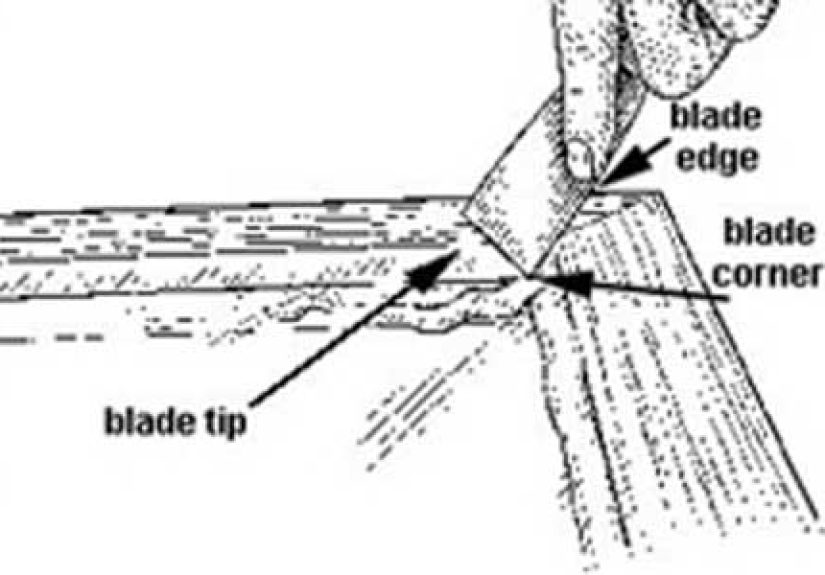
- A clean razor-cleaved edge, carbon film, or metal mesh for focus and astigmatism checks.
This gives you multiple feature sizes on one target. You can use larger mesh openings at low magnification, grid pitch at medium magnification, and smaller spheres or grating lines at higher magnification. That matters because no single feature size is ideal across every SEM magnification range. A 100-micrometer grid may be wonderful at low magnification and nearly useless at very high magnification. A 300-nanometer pattern may be excellent at high magnification and invisible when you are zoomed out.
Materials You Can Use for a DIY SEM Calibration Target
1. SEM Stub
Start with a standard aluminum SEM pin stub that fits your microscope stage. Choose the correct diameter and pin style for your instrument. A flat-top stub is easiest to work with. If your lab uses indexed or pre-labeled stubs, even better. Calibration becomes less mysterious when the target does not look like every other mysterious silver button in the drawer.
2. Conductive Carbon Tape or Conductive Adhesive
Use conductive carbon tape to hold the components in place and provide a path to ground. Poor grounding causes charging, and charging causes image drift, brightness flashes, weird shadows, and operator sadness. Apply the tape smoothly, avoid wrinkles, and use only enough adhesive to secure the sample.
3. Copper TEM Grid
A copper TEM grid can serve as a convenient low-to-medium magnification reference. Common mesh grids have repeated openings and bars, and while they are not always precise enough for formal calibration, they are useful for orientation, training, and distortion checks. They also look satisfyingly scientific, which is not a calibration metric but does help morale.
4. Commercial Grating Replica
A grating replica with known line spacing is one of the most useful pieces in a budget calibration setup. A common TEM-style calibration replica may have thousands of lines per millimeter, giving a known pitch that can be measured in the SEM or TEM. These replicas are not always as robust as silicon standards, so handle them gently and store them carefully.
5. Silicon Calibration Chip or Patterned Wafer Piece
If you can obtain a small silicon calibration chip, patterned wafer fragment, or surplus lithographic grid with known pitch, it becomes the strongest part of your DIY target. Silicon is vacuum compatible, stable, and friendly to SEM imaging. Patterned silicon grids with 1 micrometer, 10 micrometer, and 100 micrometer pitch are especially practical because they cover a useful magnification range.
6. Microspheres or Nanoparticles
Latex, silica, or polystyrene spheres with known nominal diameter can help check high-magnification behavior. However, use them carefully. Spheres may vary in size, clump together, deform under coating, or shift if mounted poorly. They are better as a secondary check than as your only calibration reference.
7. Fine Metal Mesh
Fine copper, nickel, or gold mesh can help with low-magnification calibration and distortion checks. It is also useful for seeing whether the image stretches differently in the X and Y directions. If squares look like rectangles and circles look like sad eggs, the microscope may need scan calibration or service attention.
How to Build a Practical DIY Calibration Target
Step 1: Choose Your Calibration Ranges
Before mounting anything, decide what magnification ranges you want to verify. A general SEM target should include features for low, medium, and high magnification. For example, use a 100-micrometer feature for low magnification, a 10-micrometer feature for medium magnification, and a 1-micrometer or submicrometer feature for high magnification.
If your work involves large particles, fibers, insects, coatings, or fracture surfaces, low and medium magnification may matter most. If you measure thin films, nanoparticles, microelectronic features, or surface textures, you need smaller reference features.
Step 2: Clean the Stub and Components
Cleanliness is not glamorous, but it is the difference between a calibration target and a tiny crime scene. Use clean tweezers, powder-free gloves, and a dust-free surface. Avoid touching the calibration surface. Remove loose dust with clean, dry gas if your lab allows it. Make sure the stub and components are dry before loading them into the microscope.
Do not use aggressive cleaning chemicals on delicate calibration patterns unless the manufacturer specifically recommends them. Abrasive cleaning can change line edges. Corrosive cleaning can damage surfaces. In calibration, “mostly fine” is how small errors sneak in wearing lab coats.
Step 3: Arrange the Components Like a Map
Place the largest feature near one side of the stub, the medium feature near the center, and the smallest feature on the opposite side. Leave enough space between components so you can navigate easily at low magnification. A target that requires a treasure map is not charming; it is inefficient.
A simple layout might look like this:
- Left side: copper mesh or TEM grid for low magnification.
- Center: silicon grid or patterned chip for medium magnification.
- Right side: grating replica or microspheres for high magnification.
Make a sketch or take an optical photo of the mounted target. Label the target with a unique ID, date, component list, known dimensions, and storage instructions. Future-you will appreciate this. Future-you has already misplaced three things today.
Step 4: Ground Everything
Electrical grounding is essential for stable SEM imaging. Make sure conductive components touch conductive tape or adhesive. If a component is partly insulated, add a tiny bridge of conductive paint from the edge of the sample to the stub. Do not cover the actual reference pattern. The goal is to ground the target, not bury the evidence.
Step 5: Coat Only If Necessary
Many silicon and metal calibration components are already conductive enough for SEM imaging. Nonconductive particles or polymer spheres may need a thin conductive coating, such as carbon, gold, gold-palladium, or platinum, depending on your instrument and analysis goals.
Be cautious: coating adds thickness. A heavy coating can blur fine features or change apparent particle diameter. If the target is used for EDS calibration or elemental analysis practice, carbon coating is often preferred over metal coating because metal coatings can add unwanted peaks. For simple imaging checks, a very thin sputter coating may be acceptable when charging is a problem.
How to Use Your DIY Calibration Target
Start With Stable SEM Conditions
Calibration checks should be performed under consistent conditions. Record the accelerating voltage, working distance, aperture, spot size, detector, scan speed, image resolution, chamber pressure mode, and magnification. These settings influence image quality and measurement confidence. If you change everything at once, your calibration log becomes a soup recipe.
Focus, Stigmation, and Working Distance Matter
Before measuring anything, focus carefully and correct astigmatism. A poorly focused line edge can shift the apparent boundary. A distorted beam can make circles look stretched. Use a consistent working distance, especially if your lab’s calibration protocol recommends one.
Measure Pitch, Not Just Width
For line grids and gratings, measure pitch whenever possible. Pitch is the distance from one repeated feature to the next, such as center-to-center spacing or left-edge-to-left-edge spacing. Measuring pitch across many repeated spaces reduces error compared with measuring one tiny line. For example, measuring across 20 grid spaces and dividing by 20 is usually more reliable than measuring one space and hoping the pixels behave.
Check X and Y Directions
Do not measure only horizontally. Check both X and Y directions. SEM scan distortion can differ between axes, especially if scan calibration, scan rotation, or image correction settings are off. A square grid is perfect for this because it can reveal stretching, skew, or nonlinearity.
Measure Near the Center and Edges
For serious verification, measure features near the center of the field and near the edges. Some distortion is field dependent. The center may look accurate while the outer region quietly misbehaves. This is especially important when measuring large particles or using wide fields of view.
Simple Calibration Math
The basic idea is straightforward. Compare the known feature size to the measured feature size in the SEM image. If a grid pitch is known to be 10 micrometers and the image software reports 9.8 micrometers, the image measurement is about 2% low. A correction factor can be calculated by dividing the known size by the measured size.
For example:
- Known pitch: 10.00 micrometers
- Measured pitch: 9.80 micrometers
- Correction factor: 10.00 / 9.80 = 1.0204
That means measurements taken under the same conditions would be multiplied by about 1.0204 to estimate the corrected value. However, do not blindly apply one correction factor to every magnification and setting. Calibration can vary with magnification range, scan mode, working distance, and instrument condition.
Documentation: The Secret Ingredient Nobody Brags About
A DIY calibration target is only as useful as its documentation. Keep a calibration log with the target ID, date, operator, instrument, settings, known reference dimensions, measured dimensions, correction factor, notes, and image file names. Save representative images with scale bars and raw metadata.
Good documentation turns a casual target into a repeatable tool. Bad documentation turns it into a shiny object that lives in a box and occasionally gets blamed for things.
What a DIY Calibration Target Cannot Do
A DIY calibration target is not automatically traceable, certified, or acceptable for regulated work. It may not have a known uncertainty budget. Its components may shift, age, contaminate, charge, or become damaged. If you mount a reference component crooked, measure it under poor focus, or store it uncovered in a dusty drawer, the target will not magically become accurate because you used the word “calibration.”
Use certified standards when:
- You need traceable measurements.
- Your lab follows ISO/IEC 17025 or similar quality systems.
- The results support product release, legal claims, or regulated submissions.
- You are publishing quantitative nanoscale measurements.
- You need a known uncertainty value.
For best results, treat your DIY target as a daily confidence check and use a certified standard for formal calibration. That combination is practical, affordable, and scientifically honest.
Common Mistakes to Avoid
Using Random Objects as Calibration Standards
A human hair, dust particle, insect wing, salt crystal, or mysterious crumb from the lab bench is not a calibration standard. It may be interesting. It may be beautiful. It may even look like an alien landscape. But unless its dimensions are known and stable, it cannot verify magnification.
Ignoring Charging
If the image drifts, flashes, blooms, or changes brightness during scanning, charging may be affecting the measurement. Improve grounding, lower accelerating voltage, reduce beam current, use faster scans, or apply a suitable coating when appropriate.
Measuring Too Few Features
One measurement is a hint. Several measurements are evidence. Measure multiple repeats, average the results, and record the spread. If the numbers vary wildly, the issue may be focus, contamination, distortion, or measurement technique.
Forgetting About Pixel Resolution
If a feature spans only a few pixels, the measurement will be weak. Increase image resolution or magnification so the feature is represented by enough pixels. Pixel size matters. A scale bar is not a magic wand; it is math wearing a tiny hat.
Using Damaged or Dirty Targets
Contamination can change edges, hide lines, and create misleading contrast. Store the target in a clean container, vacuum desiccator, or covered stub box. Avoid touching the surface. Retire the target if the reference area becomes scratched, coated with debris, or visibly altered.
DIY Calibration Target Ideas by Budget
Low Budget
Use a clean SEM stub, conductive carbon tape, a copper TEM grid, and a line grating replica. This setup is affordable and excellent for teaching, navigation, and basic magnification checks. It is not ideal for certified measurement, but it is far better than guessing.
Medium Budget
Add a patterned silicon chip or traceable grid standard with 1 micrometer, 10 micrometer, and 100 micrometer pitch. This provides a stronger reference for routine SEM verification and makes the target more useful across magnification ranges.
Higher Budget
Use a certified SEM magnification standard mounted on a dedicated stub, then add secondary features such as mesh, particles, or grating replicas for training and troubleshooting. Keep the certified reference area protected and avoid unnecessary coating.
Best Practices for Long-Term Use
Store the target clean, dry, and covered. Keep a digital folder with the target map, component specifications, images, and calibration log. Use the same imaging conditions for routine checks. Compare new results to previous results, not just to the nominal reference value. If the same target suddenly measures differently, investigate before trusting the next batch of measurements.
Also, create a replacement plan. DIY targets are consumables. Adhesives age. Particles detach. Coatings accumulate. Grids bend. The universe increases entropy, and electron microscopy is not exempt.
DIY Calibration Target for TEM: A Quick Note
For TEM, calibration often uses cross grating replicas, lattice fringes, nanoparticles, or known crystal spacings depending on the magnification and measurement purpose. A DIY TEM calibration setup usually means preparing a reliable grid holder and using known calibration grids or replicas, not fabricating your own nanoscale reference. TEM calibration can be more demanding because camera length, magnification, diffraction mode, and digital pixel calibration all matter.
If your work involves cryo-EM, atomic-resolution imaging, or crystallographic measurement, use appropriate standards and established lab protocols. A homemade target may help with practice and quick checks, but formal calibration belongs to validated reference materials and documented workflows.
Real-World Experience: What You Learn After Building One
The first thing you learn from building a DIY calibration target is that mounting matters more than expected. A beautiful reference grid mounted at a slight tilt, half buried in carbon tape, and decorated with dust will produce disappointing images. The SEM does not care that you had good intentions. It only sees geometry, electrons, and your mounting choices.
One practical experience is that a multi-feature target saves time. Instead of loading one standard for low magnification, another for medium magnification, and another for high magnification, a combined target lets the operator move quickly from one region to another. This is especially useful in shared labs where instrument time is precious and everyone is pretending not to watch the clock.
Another lesson is that low magnification checks are often neglected. Many users obsess over nanoscale features but forget that distortion at low magnification can affect particle counting, fiber length measurement, coating thickness overview images, and failure-analysis documentation. A simple mesh or grid pattern can reveal whether the image is stretched or skewed before serious measurement begins.
High magnification checks teach humility. A submicrometer grating or small sphere cluster will quickly expose poor focus, astigmatism, vibration, contamination, and beam damage. If the feature edges look soft, do not immediately blame the calibration target. Check focus, working distance, aperture alignment, scan speed, charging, and contamination. The target is often the messenger, not the villain.
Users also learn that target storage is not a minor detail. A target left uncovered near sample-preparation work can collect dust, fibers, adhesive residue, and mystery particles. Under the SEM, those tiny contaminants become giant scenic boulders. A clean storage box or vacuum desiccator can dramatically extend target life.
Documentation becomes more valuable over time. The first calibration log may feel excessive, but after six months it becomes a powerful troubleshooting record. If the same 10-micrometer grid measured 10.02 micrometers for months and suddenly measures 10.45 micrometers after a service visit, you have evidence worth investigating. Without the log, you only have vibes. Vibes are not metrology.
One of the best uses for a DIY calibration target is training new SEM users. Beginners often trust scale bars automatically. When they measure a known grid and see small differences caused by focus, scan settings, or measurement technique, they understand that microscopy is not just “zoom and screenshot.” It is measurement science, and measurement science likes receipts.
A DIY target also encourages better habits. Operators become more careful about grounding, sample cleanliness, beam conditions, and image metadata. They stop treating calibration as a mysterious annual ritual performed by someone with a clipboard. Instead, they see it as a routine quality check that protects every measurement that follows.
The biggest practical lesson is balance. A DIY calibration target is not a replacement for a certified standard, but it is not a toy either. Used properly, it becomes a daily confidence tool, a teaching aid, a troubleshooting sample, and a reminder that scale bars deserve respect. In electron microscopy, the difference between “looks right” and “is right” can be very small. Conveniently, that is exactly what calibration targets are built to catch.
Conclusion
A DIY calibration target for electron microscopes is a smart, affordable way to improve measurement confidence, train users, and monitor SEM or TEM performance between formal calibrations. The best version is not a risky homemade nanofabrication experiment. It is a carefully assembled, well-documented target that combines known reference features, clean mounting, good grounding, and repeatable imaging conditions.
Use it to check scale bars, compare magnification ranges, spot distortion, and build better microscopy habits. For official metrology, traceable standards still win. But for everyday confidence, a thoughtfully built DIY target can become one of the most useful small tools in the microscopy lab. It is humble, practical, and much cheaper than discovering a scale error after all your images are already in the report.