Table of Contents >> Show >> Hide
- What Is a Hemangioma of the Skin?
- What Causes Hemangioma of the Skin?
- How to Identify a Hemangioma on the Skin
- How Doctors Diagnose a Skin Hemangioma
- When a Hemangioma Needs Prompt Evaluation
- Complications Doctors Watch For
- Conditions That Can Be Mistaken for a Hemangioma
- What Happens After Diagnosis?
- Experiences People Commonly Have With Skin Hemangiomas
- Conclusion
- SEO Tags
Seeing a new red, purple, or bluish mark on the skin can send anyone into a dramatic internal monologue. Is it serious? Is it dangerous? Is Google about to ruin my evening? When the spot turns out to be a skin hemangioma, the good news is that it is usually benign. The slightly less convenient news is that “usually benign” does not mean “always ignore it.”
A hemangioma of the skin is a growth made up of extra blood vessels. It is most commonly discussed in babies, especially as infantile hemangioma, but related vascular growths can show up in other forms too. Some stay small and fade quietly. Others grow fast, ulcerate, or pop up in places where even a tiny bump can cause outsized trouble, such as the eyelid, lip, nose, or diaper area.
This guide breaks down what causes a skin hemangioma, how to identify one, how doctors diagnose it, and when a harmless-looking “strawberry mark” deserves faster medical attention. In other words, this is the calm, useful version of the internet rabbit hole.
What Is a Hemangioma of the Skin?
A skin hemangioma is a noncancerous vascular growth formed by a cluster of blood vessels. In infants, it is the most common benign tumor of the skin. Many appear shortly after birth, grow for a period of time, and then slowly shrink over the following years.
Doctors usually divide skin hemangiomas into a few practical categories:
Infantile Hemangioma
This is the classic one most people mean. It may be absent at birth or show up as a faint patch, then become more obvious within the first days to weeks of life. Infantile hemangiomas typically go through a growth phase followed by a slow involution phase, meaning they gradually flatten and fade over time.
Congenital Hemangioma
This type is already fully formed at birth. Unlike infantile hemangiomas, congenital hemangiomas do not keep growing after birth in the same way. Some shrink quickly, some shrink partially, and some do not shrink much at all.
By Depth in the Skin
Hemangiomas are also described by where they sit:
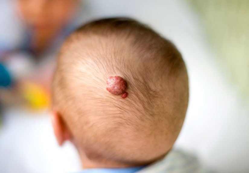
- Superficial hemangioma: bright red, raised, often called a “strawberry” hemangioma.
- Deep hemangioma: deeper under the skin, often blue, purple, or skin-colored with a swollen look.
- Mixed hemangioma: has both superficial and deep components.
That last category is a classic overachiever. It wants to be two things at once and often succeeds.
What Causes Hemangioma of the Skin?
The exact causes of hemangioma are not fully understood. The most accepted explanation is that certain blood vessels form and multiply abnormally. In plain English, the body’s tiny vessel-building crew gets a little too enthusiastic.
What experts do know is that hemangiomas are not cancer, and they are usually not caused by anything a parent did or did not do during pregnancy. There is no evidence that a specific food, activity, or everyday prenatal mistake creates them. That is an important point, because guilt tends to show up uninvited whenever a baby’s skin does something unexpected.
Several risk factors are associated with infantile hemangiomas:
- Female sex
- Premature birth
- Low birth weight
- Fair or lighter skin tone
- Multiple gestation, such as twins or triplets
Location also matters. A large share of hemangiomas appear on the head and neck, though they can develop anywhere on the body, including the trunk, arms, legs, scalp, chest, and back.
How to Identify a Hemangioma on the Skin
Hemangioma identification starts with appearance, timing, and growth pattern. Those three clues do a lot of the diagnostic heavy lifting.
Common Signs of a Superficial Hemangioma
- Bright red color
- Raised or uneven surface
- Well-defined border
- Appears in the first weeks of life
- May grow quickly in the first months
These are the marks people often call “strawberry” hemangiomas. They can start as a pale or pink area and then become more vivid and textured with time.
Common Signs of a Deep Hemangioma
- Bluish, purplish, or skin-colored swelling
- Smooth surface
- Lump or fullness under the skin
- May become obvious a bit later than superficial lesions
Deep hemangiomas are trickier because they may not look like the textbook red birthmark. Instead, they can resemble swelling, bruising, or a soft-tissue lump hiding under the skin.
Mixed Hemangiomas
Mixed lesions may have a red surface stain with a deeper bluish bulk underneath. These can look dramatic, which is unfortunate because parents were already having a stressful enough week.
How Hemangiomas Change Over Time
One of the biggest clues is behavior. Infantile hemangiomas often:
- Appear at birth or within the first few weeks
- Grow most rapidly in early infancy
- Slow down and stabilize
- Gradually flatten, fade, and shrink over several years
If a lesion follows that pattern, clinicians become more confident that it is a hemangioma rather than another vascular birthmark or skin tumor.
How Doctors Diagnose a Skin Hemangioma
Hemangioma diagnosis is often more straightforward than people expect. In many cases, a doctor can diagnose it through a physical exam and a good history.
1. Medical History
The clinician will usually ask:
- When did the mark first appear?
- Was it fully present at birth or did it show up later?
- Has it grown quickly?
- Is it painful, ulcerated, bleeding, or infected?
- Is it affecting feeding, vision, breathing, hearing, or movement?
The timing of appearance matters a lot. A lesion that is fully formed at birth may point toward a congenital hemangioma or another vascular anomaly, while one that blossoms in the first weeks of life leans more toward infantile hemangioma.
2. Physical Examination
Most skin hemangiomas are diagnosed clinically. The doctor looks at the color, shape, depth, size, and location. In dermatology, this is the part where trained eyes do what search engines cannot.
A dermatologist or pediatric specialist may also use tools such as a Wood’s lamp or close visual inspection to better evaluate skin findings.
3. Imaging When the Picture Is Unclear
Imaging is not usually required for a simple, straightforward hemangioma. However, it may be recommended when:
- The diagnosis is uncertain
- The hemangioma is deep under the skin
- There are signs of structural abnormalities
- There are multiple skin hemangiomas
- The lesion is large, segmental, or located in a high-risk area
Common imaging tests include:
- Ultrasound: useful for checking depth and blood flow
- MRI: helpful when deeper extension or involvement of nearby structures is a concern
- CT scan: used less often, but may be considered in selected cases
4. Biopsy in Rare Cases
A skin biopsy is rarely needed, but it may be used when a lesion does not behave or look like a typical hemangioma. Deep hemangiomas can sometimes resemble other vascular lesions or soft tissue masses, so biopsy becomes the “let’s stop guessing” option.
When a Hemangioma Needs Prompt Evaluation
Many hemangiomas are low-risk and only need observation. But some deserve faster assessment, especially during the early months when growth can be brisk.
Doctors are more concerned when a hemangioma is:
- Near the eye, where it may affect vision
- On the nose, lip, or ear, where it may cause disfigurement or functional problems
- In the beard area or neck, which can be associated with airway involvement
- In the diaper area, armpit, or skin folds, where ulceration is more common
- Over the lower back or lumbosacral area, where underlying anomalies may be considered
- Large, rapidly growing, or segmental
- Multiple, especially five or more, because internal hemangiomas such as in the liver may be a concern
- Ulcerated, infected, painful, or bleeding repeatedly
In these situations, early referral to dermatology, a vascular anomalies clinic, or a pediatric specialist can prevent complications and reduce long-term skin changes.
Complications Doctors Watch For
Although skin hemangiomas are benign, they are not always boring. Possible complications include:
- Ulceration: skin breakdown that can be painful and prone to infection
- Scarring or textural change: especially after larger lesions shrink
- Functional problems: affecting sight, breathing, hearing, eating, or diapering
- Residual skin changes: extra skin, visible blood vessels, wrinkling, or fatty tissue
- Psychosocial impact: especially for very visible lesions on the face
Bleeding can happen, but severe bleeding from a skin hemangioma is uncommon. When there is bleeding after injury, firm pressure is often enough to control it.
Conditions That Can Be Mistaken for a Hemangioma
Not every red or blue mark is a hemangioma. The diagnosis may need to be distinguished from:
- Vascular malformations
- Port-wine stains
- Bruising
- Pyogenic granuloma
- Other soft tissue tumors or vascular lesions
This is one reason timing matters so much. Hemangiomas have a fairly recognizable life story. Other lesions do not read from the same script.
What Happens After Diagnosis?
Once a doctor confirms a skin hemangioma, the next step is usually one of two things: watchful waiting or treatment planning.
Small, uncomplicated lesions in low-risk areas are often monitored. Higher-risk hemangiomas may be treated with options such as oral propranolol, which is considered first-line therapy for infantile hemangiomas that need treatment. Some superficial lesions may respond to topical timolol. Laser therapy, wound care, or surgery may be used in selected situations, particularly for residual skin changes or ulceration.
That said, this article is about causes, identification, and diagnosis, so we will resist the temptation to turn it into a full pharmacology lecture. You’re welcome.
Experiences People Commonly Have With Skin Hemangiomas
The medical facts matter, but so do the human experiences that go with them. Families rarely describe a hemangioma as “an interesting vascular phenomenon.” They usually describe it as “the thing that appeared on my baby’s face and made me panic at 2 a.m.” That emotional side is real and worth acknowledging.
One of the most common experiences starts with confusion. A newborn has what looks like a faint pink patch, a tiny scratch-like mark, or a subtle area of discoloration. Nobody is too alarmed at first. Then, over the next few weeks, the spot becomes brighter, puffier, and more obvious. Parents often think they missed something, but that pattern is typical of infantile hemangioma. It is not uncommon for the lesion to seem like it appeared “out of nowhere,” when really it was quietly setting up shop before becoming more visible.
Another common experience is hearing different descriptions from different people. One person calls it a birthmark. Another says strawberry hemangioma. A friend says angioma. A relative says, with breathtaking confidence and zero medical training, “That happened to my cousin and it was definitely from teething,” which is not how any of this works. The terminology can be confusing, and many families feel calmer once a specialist explains exactly what kind of vascular lesion it is.
Parents of children with facial hemangiomas often talk about the emotional weight of visibility. Even when the lesion is medically harmless, being on the eyelid, nose, lip, cheek, or forehead can lead to repeated questions from strangers. Some families worry about photographs, social reactions, and future self-esteem. That concern is not shallow or silly. Visible skin conditions affect how children are seen by others and how families experience everyday life.
There is also the experience of relief after diagnosis. Many people arrive at the appointment fearing skin cancer, a dangerous tumor, or some mysterious internal illness. When the doctor says the lesion is a benign hemangioma and may simply need observation, the room tends to exhale. At the same time, families with high-risk hemangiomas often feel relief for a different reason: now they know why early treatment matters and what the plan is.
Some of the most stressful cases involve ulceration. When a hemangioma in the diaper area or a skin fold breaks down, it can become painful, ooze, or look infected. Parents often describe this phase as the moment the lesion stopped feeling “cosmetic” and started feeling medically urgent. Pain control, skin care, and follow-up suddenly become central parts of the experience.
Finally, many people are surprised by what happens after a hemangioma fades. The red color may improve, but extra skin, fine blood vessels, a scar-like texture, or fatty tissue can remain. Families sometimes expect the mark to disappear like chalk in the rain, and instead it leaves a little reminder behind. That does not mean the diagnosis was wrong. It simply means involution and perfect cosmetic erasure are not the same thing.
In short, the experience of a skin hemangioma is often a mix of fear, watchfulness, reassurance, and patience. Medicine handles the diagnosis; time often handles the rest.
Conclusion
Hemangioma of the skin is common, usually benign, and often identifiable by its timing, color, depth, and growth pattern. Most cases can be diagnosed with a physical exam and medical history alone. The main challenge is not usually whether the lesion is dangerous, but whether it is in a location or pattern that raises the risk of complications or long-term skin changes.
If the lesion appears in early infancy, grows quickly, looks bright red or bluish, and then begins to stabilize, a hemangioma becomes a strong possibility. If it is large, painful, ulcerated, multiple, or close to the eye, mouth, airway, or diaper area, earlier specialist evaluation is wise. In skin medicine, timing matters. The sooner a high-risk hemangioma is recognized, the better the chance of protecting both function and appearance.
So yes, a hemangioma can look dramatic. But with accurate identification, proper diagnosis, and the right follow-up, it is usually far less scary than it first appears.