Table of Contents >> Show >> Hide
- What Is a Mammogram?
- Screening Mammogram vs. Diagnostic Mammogram
- Why Mammograms Matter
- When Should You Start Getting Mammograms?
- Who May Need Earlier or Extra Screening?
- What Happens During a Mammogram?
- 2D Mammogram vs. 3D Mammogram
- Dense Breasts: What That Means
- Benefits of Mammograms
- Limitations and Risks of Mammograms
- How to Prepare for a Mammogram
- Understanding Mammogram Results
- When to Call Your Doctor
- Common Mammogram Myths
- Access, Cost, and Practical Concerns
- Experiences Related to Mammograms: What Real Life Often Feels Like
- Conclusion
A mammogram is one of those health appointments that can sound more intimidating than it usually is. The word itself feels like it belongs on a spaceship dashboard, and the machine is not exactly winning any spa design awards. But behind the awkward positioning, brief pressure, and “please don’t wear deodorant” instructions is a powerful breast cancer screening tool that helps detect changes in breast tissue before they can be felt.
Knowing the facts about mammograms matters because the advice can feel confusing. Some organizations recommend screening every year. Others recommend every two years. Some women need to start earlier than age 40, while others may continue after 74 depending on health and personal preference. Add dense breast tissue, 3D mammograms, false positives, and insurance questions to the mix, and suddenly a simple appointment can feel like a group project no one volunteered for.
This guide breaks it down in plain American English: what mammograms do, when to get one, what to expect, what the results mean, and how to make smart decisions with your healthcare provider.
What Is a Mammogram?
A mammogram is a low-dose X-ray image of the breast. It is used to look for signs of breast cancer, including masses, tiny calcium deposits called calcifications, and changes in breast tissue patterns. Mammograms are not perfect, but they remain one of the most important tools for early breast cancer detection.
The goal of a screening mammogram is not to diagnose cancer on the spot. Instead, it looks for changes that may need a closer look. Think of it as a smoke alarm, not a fire investigation team. If something looks unusual, your provider may recommend additional imaging, such as a diagnostic mammogram, breast ultrasound, MRI, or biopsy.
Screening Mammogram vs. Diagnostic Mammogram
Screening mammogram
A screening mammogram is done when there are no breast symptoms. It is a routine check designed to find breast cancer early, often before a lump can be felt. This is the type most people mean when they say, “I’m due for my mammogram.”
Diagnostic mammogram
A diagnostic mammogram is used when there is a specific concern. That concern may be a lump, nipple discharge, breast pain in one area, skin changes, a previous abnormal screening result, or a need to take extra views of a certain area. Diagnostic mammograms usually take longer than screening mammograms because the radiology team may capture more detailed images.
Why Mammograms Matter
Breast cancer is easier to treat when found early. Mammograms can detect some cancers before they cause symptoms, which may allow treatment to begin at an earlier stage. Early detection can also mean more treatment options, less extensive surgery for some patients, and a better chance of long-term survival.
That said, a mammogram is not a magic crystal ball. It can miss some cancers, especially in people with dense breast tissue. It can also find suspicious areas that turn out not to be cancer. The smartest approach is not blind panic or blind trust; it is informed screening, regular follow-up, and a plan based on your personal risk.
When Should You Start Getting Mammograms?
For women at average risk, many major U.S. medical organizations now support starting breast cancer screening at age 40, but they differ on how often mammograms should be done.
- U.S. Preventive Services Task Force: Recommends mammograms every two years for women ages 40 to 74.
- American Cancer Society: Says women ages 40 to 44 should have the option to start yearly screening; women ages 45 to 54 should get mammograms every year; women 55 and older may switch to every other year or continue yearly screening.
- American College of Obstetricians and Gynecologists: Recommends that average-risk individuals be offered screening starting at age 40, with shared decision-making about frequency.
- American College of Radiology and Society of Breast Imaging: Recommend annual mammograms beginning at age 40 for average-risk women.
- NCCN guidance: Generally supports annual mammography with tomosynthesis starting at age 40 for average-risk women.
So, which schedule is “right”? The best answer depends on your risk factors, values, health history, and comfort with the benefits and downsides of screening. If you are 40 or older, the key move is to talk with your healthcare provider and choose a schedule you can realistically keep.
Who May Need Earlier or Extra Screening?
Some people have a higher-than-average risk of breast cancer and may need mammograms earlier than age 40 or additional screening with breast MRI or ultrasound. Higher-risk factors may include:
- A BRCA1, BRCA2, PALB2, TP53, or other inherited gene mutation linked to breast cancer
- A strong family history of breast cancer, especially in a parent, sibling, or child
- Previous chest radiation therapy at a young age
- A personal history of breast cancer
- Certain high-risk breast biopsy findings, such as atypical hyperplasia or lobular carcinoma in situ
- A calculated lifetime breast cancer risk of 20% or higher
If any of these apply, do not wait for a generic screening reminder postcard to make decisions for you. Ask your provider about formal breast cancer risk assessment and whether genetic counseling, MRI screening, or a personalized plan makes sense.
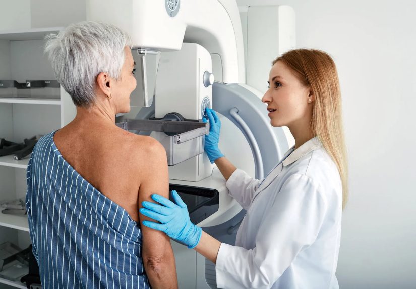
What Happens During a Mammogram?
A mammogram appointment is usually quick. The imaging itself often takes only a few minutes, though the full visit may take longer because of check-in, changing clothes, and positioning.
You will typically undress from the waist up and put on a gown. A technologist will position one breast at a time on the mammography machine. The breast is gently but firmly compressed between two plates while images are taken. Compression can feel uncomfortable, but it helps spread the tissue, reduce motion, improve image quality, and lower the amount of radiation needed.
Yes, the compression is awkward. No, it is not supposed to feel like a medieval punishment device. If the pain is intense, tell the technologist immediately. They can usually adjust positioning.
2D Mammogram vs. 3D Mammogram
A traditional mammogram creates two-dimensional images of the breast. A 3D mammogram, also called digital breast tomosynthesis, takes multiple images from different angles and creates a layered view of breast tissue. Many facilities now offer 3D mammography, and it may be especially helpful for some people with dense breasts.
Both 2D and 3D mammograms use low-dose X-rays. A 3D mammogram may slightly increase radiation exposure compared with 2D alone, but the dose remains low and within regulated safety limits. It may also reduce the chance of being called back for extra images in some patients, because radiologists can examine breast tissue layer by layer instead of trying to read the entire “sandwich” at once.
Dense Breasts: What That Means
Breasts are made of fatty tissue, glandular tissue, and fibrous tissue. Dense breasts have more glandular and fibrous tissue compared with fatty tissue. Breast density is common and cannot be determined by touch, breast size, or firmness. Only a mammogram can show whether breast tissue is dense.
Dense breast tissue matters for two reasons. First, it can make cancer harder to see on a mammogram because both dense tissue and many tumors appear white on the image. Second, dense breasts are linked with a higher risk of developing breast cancer.
Since September 2024, mammography facilities in the United States have been required to notify patients about breast density in mammogram results. If your letter says you have dense breasts, do not panic. Dense breasts are not rare, and they do not mean you have cancer. They do mean you should ask your provider whether your overall risk calls for 3D mammography, ultrasound, MRI, or a different screening schedule.
Benefits of Mammograms
The biggest benefit of mammography is early detection. Mammograms can find breast cancers before they are large enough to feel. They can also detect ductal carcinoma in situ, or DCIS, which is abnormal cell growth inside a milk duct. Some DCIS may become invasive cancer, while some may not progress. This is one reason screening decisions involve both benefits and trade-offs.
Regular mammograms also create a record over time. Comparing new images with older ones can help radiologists identify subtle changes. That is why it is helpful to return to the same imaging center when possible or bring prior images if you switch facilities.
Limitations and Risks of Mammograms
False positives
A false positive happens when a mammogram finds something suspicious that later turns out not to be cancer. This can lead to more imaging, short-term follow-up, or biopsy. False positives can be stressful, expensive, and inconvenient. The emotional roller coaster is real, and no one enjoys being told, “We need more pictures,” especially when your brain immediately starts writing a disaster movie.
False negatives
A false negative means a mammogram looks normal even though cancer is present. This is more likely in people with dense breast tissue. That is why new symptoms should never be ignored just because a recent mammogram was normal.
Overdiagnosis
Overdiagnosis means finding a cancer that would not have caused symptoms or death during a person’s lifetime. Because doctors cannot always predict which cancers will become dangerous, treatment may still be recommended. This is one of the reasons experts continue to debate the ideal screening schedule.
Radiation exposure
Mammograms use a low dose of radiation. For most people, the benefit of screening outweighs this small risk. If you are pregnant, think you might be pregnant, or are breastfeeding, tell your healthcare team before the exam.
How to Prepare for a Mammogram
A little preparation can make the appointment smoother:
- Schedule the exam for a time when your breasts are less likely to be tender, often the week after your period if you menstruate.
- Do not wear deodorant, antiperspirant, lotion, powder, or perfume under your arms or on your chest. These can show up as spots on the image.
- Wear a two-piece outfit so you only need to remove your top.
- Bring prior mammogram records if you are visiting a new facility.
- Tell the technologist about breast implants, prior surgery, hormone therapy, or any new symptoms.
- Ask when and how you will receive results.
Understanding Mammogram Results
Mammogram reports often use BI-RADS, which stands for Breast Imaging Reporting and Data System. It is a standardized way radiologists describe findings and follow-up recommendations.
- BI-RADS 0: More imaging is needed.
- BI-RADS 1: Negative; no concerning finding.
- BI-RADS 2: Benign finding.
- BI-RADS 3: Probably benign; short-term follow-up may be recommended.
- BI-RADS 4: Suspicious abnormality; biopsy may be recommended.
- BI-RADS 5: Highly suggestive of cancer; appropriate action is needed.
- BI-RADS 6: Known biopsy-proven cancer.
If your result says you need more imaging, it does not automatically mean cancer. Many callbacks happen because the radiologist needs a clearer view or wants to compare a specific area more carefully.
When to Call Your Doctor
Do not wait for your next scheduled mammogram if you notice a new breast change. Contact your healthcare provider if you have:
- A new lump or thickened area in the breast or underarm
- Nipple discharge, especially if bloody or one-sided
- New nipple inversion
- Skin dimpling, redness, scaling, or thickening
- Persistent breast pain in one specific area
- A change in breast size or shape that is new for you
Most breast changes are not cancer, but they deserve attention. Your body is allowed to be dramatic; your job is to get the plot checked.
Common Mammogram Myths
Myth: Mammograms cause breast cancer.
Fact: Mammograms use low-dose radiation. The risk from radiation exposure is very small for most people, while the benefit of early detection can be significant.
Myth: If I have no family history, I do not need screening.
Fact: Many people diagnosed with breast cancer do not have a strong family history. Family history matters, but it is not the whole story.
Myth: A normal mammogram means I can ignore symptoms.
Fact: No screening test is perfect. New symptoms should be evaluated even after a normal mammogram.
Myth: Dense breasts are abnormal.
Fact: Dense breasts are common. They are not a disease, but they can affect screening accuracy and breast cancer risk.
Access, Cost, and Practical Concerns
Screening mammograms are often covered by health insurance for eligible patients, but coverage for diagnostic imaging, ultrasound, MRI, or follow-up tests may vary. If cost is a concern, ask about hospital financial assistance, state screening programs, community clinics, and the National Breast and Cervical Cancer Early Detection Program.
When scheduling, ask whether the facility is certified under the Mammography Quality Standards Act. Certification helps ensure the facility meets federal quality standards for equipment, personnel, image quality, and reporting.
Experiences Related to Mammograms: What Real Life Often Feels Like
The facts are important, but mammograms are not lived on paper. They happen in real rooms, with paper gowns that open in confusing directions, appointment reminders that arrive at inconvenient times, and people quietly wondering whether everyone else is also nervous. Spoiler: many are.
For a first mammogram, the biggest experience is often uncertainty. A person may walk in expecting the appointment to be painful, embarrassing, or complicated. In reality, the awkwardness is usually worse than the pain. The technologist has done this many times and is focused on getting clear images, not judging anyone’s body. The positioning may feel odd because it is oddly specific: turn here, lean there, hold still, relax your shoulder, do not breathe for one second. It can feel like breast yoga directed by a very polite air traffic controller.
Many people are surprised by how fast the actual imaging is. The compression lasts only seconds for each picture, though those seconds are not exactly anyone’s favorite vacation memory. The appointment can still feel emotionally heavy because breast cancer screening carries a “what if?” feeling. Bringing a calm mindset helps. So does planning a small reward afterward: coffee, a walk, a favorite podcast, or simply crossing it off the list with the satisfaction of an adult who has defeated the calendar.
Another common experience is the callback. A callback can sound scary, but it often means the radiologist needs additional images. This is especially common after a first mammogram because there are no older images for comparison. It may also happen with dense breasts, overlapping tissue, cysts, calcifications, or a blurry view. The waiting period can be stressful. A helpful strategy is to ask direct questions: “What did you see?” “What test comes next?” “When will I get results?” Information does not remove every worry, but it does stop imagination from becoming the unpaid director of a horror film.
For people who receive a dense breast notification, the experience can be confusing. The letter may say the mammogram was normal and also say dense tissue can make cancer harder to see. That sounds like the medical version of “good news, but also please keep reading.” The practical next step is not panic; it is a conversation. Ask whether your breast density plus your age, family history, prior biopsies, genetics, and other risk factors change your screening plan.
Some people build mammograms into a yearly health routine. They schedule around a birthday month, Breast Cancer Awareness Month, or an annual physical. Others prefer every two years based on provider guidance. What matters most is having a plan, keeping track of results, and not disappearing after a false positive or uncomfortable experience. Screening works best when it becomes a habit rather than a once-in-a-decade surprise event.
There is also an emotional experience that deserves respect: fear. Some people avoid mammograms because they are afraid of pain, radiation, results, cost, or being dismissed. Those concerns are not silly. A good healthcare team should explain the process, answer questions, and help you understand your options. Mammograms are not about being fearless. They are about taking one practical step even if you are carrying a pocketful of nerves.
Conclusion
Mammograms are not perfect, but they are valuable. They can find breast cancer early, guide follow-up testing, and help create a long-term record of breast health. The key facts are simple: average-risk women should generally begin discussing routine mammography by age 40; screening frequency depends on the guideline and personal risk; dense breasts may require extra discussion; and abnormal results do not automatically mean cancer.
The smartest mammogram plan is personal, practical, and consistent. Know your risk factors, understand your results, ask about breast density, and keep the conversation going with your healthcare provider. A mammogram may be brief, awkward, and not exactly glamorous, but it is also a meaningful act of preventive care. In other words: not fun, but absolutely worth knowing about.
Note: This article is for educational purposes only and does not replace professional medical advice, diagnosis, or treatment. Readers should speak with a qualified healthcare provider about personal breast cancer risk and screening decisions.